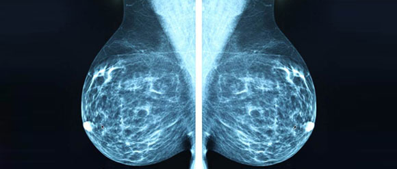
Mammography

Mammography is screening technique to diagnose malignancy in the breast. It uses low energy X rays to examine the breast. The motive of a mammography is early detection of breast cancer so as to treat it in time and cure it. Mammography followed by other screening methods such as ultrasound, PET scan, MRI and ductography aid in confirming malignancy of tumors in the breast.
While mammography aids in initial screening and diagnosis of breast lumps and cancer, it can also be misleading. A clear mammogram can indicate exact location and density of the tumor. However several times, due to many dense tissues in the breast there can be false positives and false negatives.
False positives can lead to unnecessary screening and further expenses for a negative malignancy causing unwanted stress and anxiety. On the other hand, a false negative can be more dangerous as the whole purpose of early detection is lost.
A mammography unit is used to perform a mammography. Breast is placed on the parallel plates of the mammography unit and compressed in order to even out the thickness of the breast tissue. This ensure lesser diffusion of x rays and clearer image. It is recommended to avoid applying deodorant, talcum powder or lotion before screening the breast as these appear as calcium deposits and can be misleading. There are two types of mammography or mammogram studies:
Screening Mammography : This type is generally for the general yearly screening of the breast and yields 4 standard X Ray images . This includes the craniocaudal (CC) view and the mediolateral oblique view.
Diagnostic Mammography: This type is for patients with known breast irregularities, changes, conditions or history. Apart from the standard X Ray images, geometrically magnified and spot-compressed views are also taken.
Mammography has advanced to digital mammography where digital receptors and software is used to analyse the digital mammograms instead of X Ray films to study the breast tissue. Digital mammography are of the two types: Spot View for breast biopsy and Full Field for screening.
3D mammography, also known as digital breast tomosynthesis (DBS), tomosynthesis, and 3D breast imaging, is a mammogram technology that creates a 3D image of the breast using X-rays. It adds value to a regular mammography however exposes the patient to radiation two times.


